
www.usa-automation.net
21
'20
Written on Modified on
Stemmer imaging News
Raman spectroscopy measures corona infected cells
With BioRam®, CellTool has developed a Raman microscope system that allows to identify and characterize cells without labelling – solely based on interaction of photons with biomolecules of a cell. The recorded Raman spectra are as unique as a fingerprint.
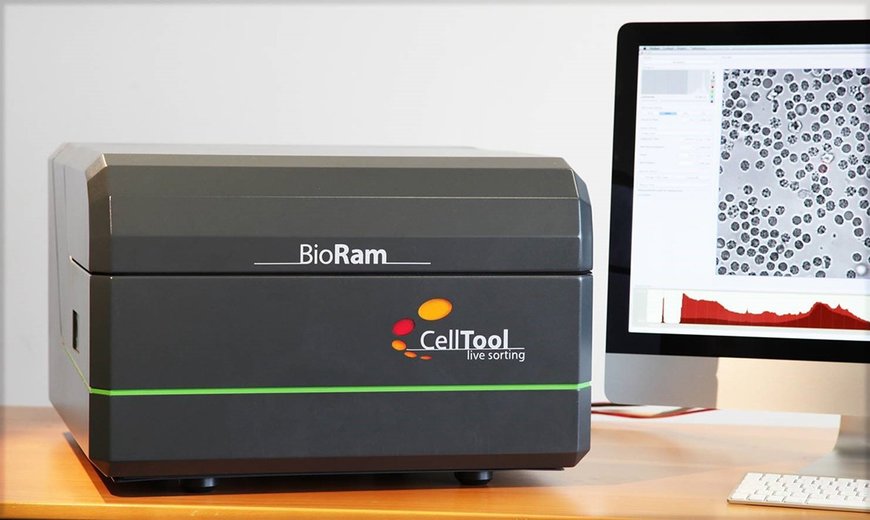
BioRam® was recently even used to identify corona-infected cells and to measure patient blood cells. A direct detection of bacteria without prior cultivation or the measurement of extracellular vesicles such as exosomes is fast and easy. Vision components from STEMMER IMAGING play an important role for the BioRam® microscope of the newly introduced second generation.
Not only related to the outbreak of the Corona pandemic cell analysis, advanced medical diagnostics or quality control of cell-based products have always been important for answering biomedical questions. However, due to the worldwide Covid-19 developments, spring 2020 highlighted the importance of progress in medical analysis in order to be able to quickly detect threatening diseases and combat them effectively.
An innovative possibility is now coming from the Bavarian town of Tutzing: the medical technology company CellTool has presented the second generation of its BioRam® Raman-Trapping-Microscope system, which is optimized for biomedical research and enables, amongst other things, the detection of diseased or infected cells, the determination of tumor aggressiveness, the discovery of biomarkers, the tracking of differences of tumor cells, the observation of the differentiation potential of stem cells or the determination of the quality of cell-based products. This Raman microscope also allows an automated analysis of blood cells to monitor their loss of functionality, e.g. due to storage time of a blood product or caused by bacterial or viral contamination.
As simple as light microscopy
"BioRam® is a highly sensitive confocal Raman trapping microscope that makes Raman spectroscopy as easy as light microscopy," said Dr. Karin Schütze, founder & CSO of CellTool. Raman spectroscopy is named after it’s inventor Sir C. V. Raman - an Indian physicist. It is based on recording the interaction of focused laser light with biomolecules of the cell. The resulting inelastic scattered light yields a sum spectrum that is characteristic for each cell type and cell state and thus, enables unique analysis of cell properties that can be used for cell diagnostics.

According to Schütze, Raman microscopy is very versatile: "Wherever antibodies and markers reach their limits or where only a small amount of sample material is available, this method offers excellent possibilities. BioRam® is an ideal system that is able to identify and characterize cells non-invasively and under physiological conditions. Thus, the cells remain vital and can be reuse in their original state."
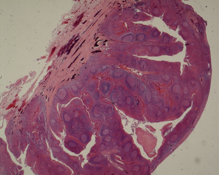
According to Schütze, living or fixed cells in cell culture or histological tissue sections can be examined just as easily as cells in 3D tissue and scaffolds. Also solids and liquids as well as cell supernatants or vaccines can be easily analyzed with BioRam®.
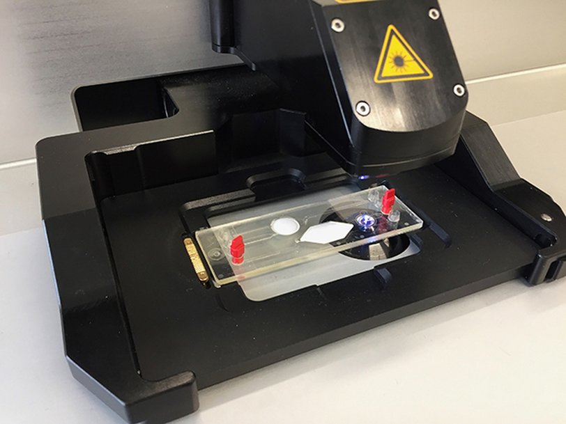
As a special feature of BioRam®: Schütze highlights the integrated optical tweezers, for which Dr. Art Ashkin was awarded a Nobel Prize in 2018. "It enables the examination of moving samples in fluids because the cells are arrested within the laser focus during Raman analysis. In addition, the optical tweezers can be used to move and reposition cells or particles".
Schütze met Art Ashkin during her post-doc time in US. She assembled her first optical trap under his supervision and together they studied the force of organelle transportation, published in Nature magazine in 1990.
Automated view into the microscope
The evaluation of microscope images is usually time-consuming and requires experienced staff. In order to make this step more economical, CellTool got in touch with the machine vision experts at STEMMER IMAGING during the development phase of the second BioRam® generation. "The advice we received exceeded our expectations," recalls Schütze: "Very fast we received the results of an extensive feasibility study carried out in STEMMER IMAGING's application laboratory, which provided us with well-founded recommendations for a suitable camera."
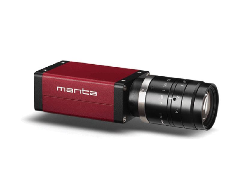
On this basis, CellTool finalized the development of the new digital BioRam® generation and integrated a Manta GigE color area scan camera from Allied Vision. The resolution of 1388x1038 pixels provided the necessary detailed information for the subsequent automatic analysis.
"Instead of a human being, a camera takes care of looking into the eyepiece and allows a much more reliable and faster analysis of the samples," says Schütze, explaining the positive result of the cooperation with STEMMER IMAGING.
Versatile application possibilities
Schütze is absolutely convinced of the performance of the new BioRam®: "Most of our results have been confirmed using common methods such as FACS, MACS, immunocytochemistry, DNA or RNA arrays. However, these methods are much more complex and require far more cell material or cannot be used for living cells as for staining cells usually have to be fixed (endpoint analysis method). In addition antibodies often lack specificity or are simply not available for certain cell types or cell states."
There are almost no limits concerning applications for the new product: Besides the analysis of biological and medical samples for research and development, BioRam® can also be used for the quality control of cell based therapeutics, but also of scaffolds, membranes, cell cultures or vaccines.
"We are sure that medical laboratories will benefit considerably from the advantages of the new BioRam® because it enables faster analysis and simple process monitoring," said Schütze. They will profit from the reduction of costs and time-consuming preparation steps and get the possibility to quickly obtain an overview of drug reactions or being able to monitor the quality of cell cultures or cell-based products. The new generation also integrates fluorescence which offers customers the possibility to view and measure fluorescence-stained samples.
"BioRam® is the ideal platform for novel cell analyses, advanced medical diagnostics or quality assurance of cell-based products. This system offers the necessary prerequisites for direct measurements in cell culture, tissue, transplants or fluids," emphasizes Schütze.
www.stemmer-imaging.com